
We provide access, training, and ongoing support for users of a wide range of optical (light) microscopes. From stereo and widefield microscopes to more advanced techniques like confocal, multiphoton, and superresolution, the facility and the managers are available to provide technical support and consultation to advance scientific inquiry and aid in experimental design.
We have instruments and support staff in two locations:
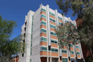
Our location in the basement of the Marley building (main campus) is equipped with two Zeiss LSM880 34 channel Laser Scanning Confocal Microscopes. One system is an inverted laser scanning confocal microscope with seven laser lines, motorized stage, and temperature control capability. The second system is an upright laser scanning confocal/multiphoton microscope with six laser lines, motorized stage, Zeiss Fast Airyscan super-resolution attachment, and temperature control as well as a Spectra Physics MaiTai femtosecond pulsed laser for multi-photon imaging. In addition, we have a Zeiss Axiozoom 16 brightfield/fluorescent stereo microscope with a Hamamatsu Flash 4.0 16bit sCMOS camera with fluorescence filters for imaging UV (DAPI), Blue (GFP, FITC, Alexa488) and Green (RFP, Alexa 555). The facility has ancillary equipment and space to support sample preparation and sample analysis for the light microscopes.
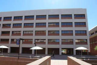
Our location in the Life Sciences North building (University of Arizona Health Sciences) includes a fully motorized, computer controlled Leica DMI6000 inverted microscope which can be used to capture images in brightfield, DIC (mags ≥20x), polarized light, or four channels of fluorescence, using either a 16bit greyscale camera or a 24bit color camera. Life Sciences North also houses a Zeiss Axio Observer 7 with Apotome III microscope, as well as a Zeiss Elyra S.1 superresolution optical microscope. The Apotome system is a multifunction widefield microscope that can give confocal-like images of samples up to 20um in thickness. The structured illumination microscope (SIM) can be used to image fluorescence markers at twice the resolving power of confocal or deconvolution microscopes. We also provide access to and support for an image analysis workstation that features Hamamatsu's HCImage software, file viewer software for the major microscope vendors, as well as other open-source image analysis tools (ImageJ/FIJI, Cell Profiler, QuPath).
Location
The core facility has two locations with typical work hours of:
Monday - Friday: 8:30AM - 5:00PM
(24/7 access for Trained Users)
Life Sciences North
(4th floor rooms 410B, 429)
1333 N Martin Ave
Tucson, AZ 85719 USA
Get in touch

Contact person
Doug Cromey, M.S.
Ved Prakash, Ph.D.
Facility Email
opticalimaging@arizona.edu
Phone
520-626-2824 (LSN)
520-621-5097 (Marley)
Reference this core in grants and publications using:
University of Arizona Imaging Cores - Optical, RRID:SCR_023355