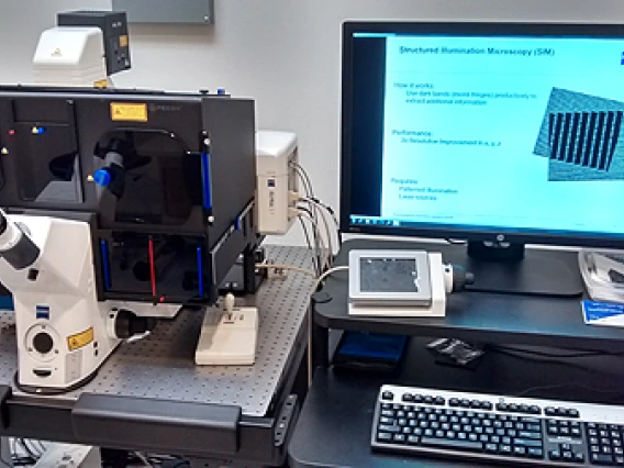
The Zeiss Elyra S.1 uses the technique of structured illumination (SR-SIM) to achieve resolutions up to two times better than that of a confocal, deconvolution or standard fluorescence microscope. Resolutions in XY of 110nm and in Z of 300nm are possible (resolution is wavelength and sample-preparation dependent), allowing users to image a volume in the sample that is 1/8 that of previous techniques.
The good news is that this super-resolution technique is a not-too-difficult jump from the dyes and sample prep techniques used for a confocal or deconvolution microscope. The Elyra S.1 can image four fluorescent colors, roughly corresponding to DAPI, FITC/GFP, Rhodamine/RFP, and CY5 (see below). SR-SIM can image to depths of 10-15 microns into a sample. Note: SR-SIM does not work well (or at all) with dimly fluorescent samples or samples that photo-bleach.
The Elyra S.1 has the following microscope objectives:
- Plan-Neofluar 10x/0.30
- Plan-APO 40x/1.4 Oil
- Plan-Apochromat 63x/1.40 Oil
- Plan-Apochromat 63x/1.30 Oil/Water/Glycerine KORR
- Alpha Plan-APO 100x/1.46 Oil
The laser lines are 405, 488, 561, and 642nm. Imaging can be performed somewhat faster using a single multi-band cube, or with 4 sequential cubes to reduce spectral overlap:
- EF LBF 405/488/561/642
- EF BP 420-480 / BP 495-550 / LP 655
- EF BP 495-550/BP 570-620
- BP 420-480/LP 655


